lv summit pvc ablation | ablation of lv summit lv summit pvc ablation Figure 3 Successful ablation of left ventricular summit (LVS) premature ventricular complex (PVC) from the basal left ventricular endocardium. Earliest activation was recorded at a unipolar wire (arrowhead) advanced into a septal venous perforator (earlier than great cardiac vein/anterior interventricular vein and any endocardial site . Need help with your product? Let us help you find what you need. Find support for your Canon LV-5220. Browse the recommended drivers, downloads, and manuals to make sure your product contains the most up-to-date software.
0 · septal vein ablation diagram
1 · pvcs for lv summit
2 · lvot ablation
3 · left ventricular summit ablation
4 · left ventricular arrhythmia ablation
5 · left ventricular ablation diagram
6 · left ventricular ablation
7 · ablation of lv summit
Tetris is one of the most classic examples of the "easy to learn difficult to master" design principle. To help you get better at Tetris, we asked Jonas Neubauer, seven-time winner of the Classic .
Figure 3 Successful ablation of left ventricular summit (LVS) premature ventricular complex (PVC) from the basal left ventricular endocardium. Earliest activation was recorded at . Premature ventricular contractions originating from the left ventricular (LV) summit pose a serious challenge to catheter ablation, as . Figure 3 Successful ablation of left ventricular summit (LVS) premature ventricular complex (PVC) from the basal left ventricular endocardium. Earliest activation was recorded at a unipolar wire (arrowhead) advanced into a septal venous perforator (earlier than great cardiac vein/anterior interventricular vein and any endocardial site . Premature ventricular contractions originating from the left ventricular (LV) summit pose a serious challenge to catheter ablation, as myocardial thickness, epicardial fat, and coronary vessels impede appropriate radiofrequency (RF) energy delivery to the target areas.
LV summit VAs are most commonly ablated within the GCV or AIVV but sometimes from the epicardial surface more lateral to these venous structures. Several LV summit VAs arise from an area that is inaccessible to catheter ablation, bounded by the left coronary, arteries and superior to the GCV and AIVV. ECG algorithms have also been developed to assist in distinguishing LV summit site that are outside the ablation inaccessible zone and within the more lateral accessible zone . An RBBB, TZ 1.1 and S wave in V 5 or V 6 predicted whether an LV summit OTVA could be ablated in the accessible area. 35 Ablation of LVS arrhythmias may be performed at the septal right ventricular outflow tract (RVOT), the left coronary cusp, the LV myocardium beneath the left coronary cusp, the distal coronary sinus, and the great cardiac vein, as well as via the epicardial approach. We performed stepwise catheter ablation on the LV-summit PVC origin site adjacent to severe coronary artery stenosis using a 3D electroanatomic mapping system in a single case. Special precautions should be taken to avoid coronary artery damage during ablation from distal CVS.
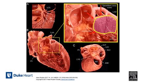
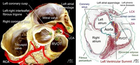
While the overall success rate of premature ventricular contraction (PVC) ablation approaches 85%, 1 ventricular arrhythmia (VA) from the left ventricular (LV) ostium and LV summit poses particular challenges with higher rates of ablation failure. 2,3 Accurate localization of the LV ostium sites of origin and an improved understanding of the opt.Surgical cryoablation in the LV summit is a viable option for drug-refractory ventricular arrhythmias. Presurgical epicardial mapping can facilitate the surgical procedure by localizing the area of interest to allow for a more limited surgical dissection of the epicardial fat.LV VAs.2 The complex relationships between the left ventricular summit (LVS) and surrounding structures under-score the importance of understanding the anatomy of this region and the value of imaging techniques for detailed mapping and safe ablation. In this article, we review the anatomy of the LVS and our approach to mapping and
Premature ventricular contractions originating from the left ventricular (LV) summit pose a serious challenge to catheter ablation, as myocardial thickness, epicardial fat, and coronary vessels impede appropriate radiofrequency (RF) energy delivery to the target areas. Figure 3 Successful ablation of left ventricular summit (LVS) premature ventricular complex (PVC) from the basal left ventricular endocardium. Earliest activation was recorded at a unipolar wire (arrowhead) advanced into a septal venous perforator (earlier than great cardiac vein/anterior interventricular vein and any endocardial site . Premature ventricular contractions originating from the left ventricular (LV) summit pose a serious challenge to catheter ablation, as myocardial thickness, epicardial fat, and coronary vessels impede appropriate radiofrequency (RF) energy delivery to the target areas.
LV summit VAs are most commonly ablated within the GCV or AIVV but sometimes from the epicardial surface more lateral to these venous structures. Several LV summit VAs arise from an area that is inaccessible to catheter ablation, bounded by the left coronary, arteries and superior to the GCV and AIVV. ECG algorithms have also been developed to assist in distinguishing LV summit site that are outside the ablation inaccessible zone and within the more lateral accessible zone . An RBBB, TZ 1.1 and S wave in V 5 or V 6 predicted whether an LV summit OTVA could be ablated in the accessible area. 35 Ablation of LVS arrhythmias may be performed at the septal right ventricular outflow tract (RVOT), the left coronary cusp, the LV myocardium beneath the left coronary cusp, the distal coronary sinus, and the great cardiac vein, as well as via the epicardial approach. We performed stepwise catheter ablation on the LV-summit PVC origin site adjacent to severe coronary artery stenosis using a 3D electroanatomic mapping system in a single case. Special precautions should be taken to avoid coronary artery damage during ablation from distal CVS.

septal vein ablation diagram
While the overall success rate of premature ventricular contraction (PVC) ablation approaches 85%, 1 ventricular arrhythmia (VA) from the left ventricular (LV) ostium and LV summit poses particular challenges with higher rates of ablation failure. 2,3 Accurate localization of the LV ostium sites of origin and an improved understanding of the opt.Surgical cryoablation in the LV summit is a viable option for drug-refractory ventricular arrhythmias. Presurgical epicardial mapping can facilitate the surgical procedure by localizing the area of interest to allow for a more limited surgical dissection of the epicardial fat.LV VAs.2 The complex relationships between the left ventricular summit (LVS) and surrounding structures under-score the importance of understanding the anatomy of this region and the value of imaging techniques for detailed mapping and safe ablation. In this article, we review the anatomy of the LVS and our approach to mapping and


rolex 5678 band

pvcs for lv summit
View and Download Canon LV-7575 specifications online. LV-7575 EXPAND SERIAL COMMAND FUNCTIONAL SPECIFICATIONS. LV-7575 projector pdf manual download. Also for: Lv-x4.
lv summit pvc ablation|ablation of lv summit


